Pages

Friday, January 25, 2013

Cancer Stages


Posted By: 

Cancer staging is the action of free the admeasurement to which a blight has developed by spreading. Contemporary convenance is to accredit a amount from I-IV to a cancer, with I getting an abandoned blight and IV getting a blight which has advance to the absolute of what the appraisal measures. The date about takes into annual the admeasurement of a tumor, how acutely it has penetrated aural the bank of a alveolate agency (intestine, urinary bladder), whether it has invaded adjoining organs, how abounding bounded lymph nodes it has metastasized to (if any), and whether it has advance to abroad organs.

Points:


  • Staging systems for cancer have evolved over time and continue to change as scientists learn more about cancer.
  • Staging describes the extent or severity of a person’s cancer. Knowing the stage of disease helps the doctor plan treatment and estimate the person’s prognosis.
  • Most tumors can be described as stage 0, stage I, stage II, stage III, or stage IV.
  • The TNM staging system is based on the extent of the tumor (T), whether cancer cells have spread to nearby (regional) lymph nodes (N), and whether distant (to other parts of the body) metastasis (M) has occurred.
  • Physical exams, imaging procedures, laboratory tests, pathology reports, and surgical reports provide information to determine the stage of the cancer. 

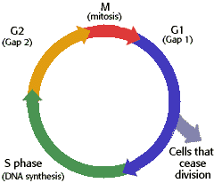
Staging helps the doctor plan the appropriate treatment. The stage can be used to estimate the person’s prognosis. Staging helps health care providers and researchers exchange information about patients; it also gives them a common terminology for evaluating the results of clinical trials and comparing the results of different trials. Knowing the stage is important in identifying clinical trials that may be suitable for a particular patient. Staging is based on knowledge of the way cancer progresses. Cancer cells grow and divide without control or order, and they do not die when they should. As a result, they often form a mass of tissue called a tumor. As the tumor grows, it can invade nearby tissues and organs. Cancer cells can also break away from the tumor and enter the bloodstream or the lymphatic system. By moving through the bloodstream or lymphatic system, cancer cells can spread from the primary site to lymph nodes or to other organs, where they may form new tumors. The spread of cancer is called metastasis.

Stage
Definition
Stage 0Carcinoma in situ.
Stage I, Stage II, and Stage IIIHigher numbers indicate more extensive disease: Larger tumor size and/or spread of the cancer beyond the organ in which it first developed to nearby lymph nodes and/or organs adjacent to the location of the primary tumor.
Stage IVThe cancer has spread to another organ(s).


TNM Staging  System

'TNM' stands for Tumour, Node, Metastasis. The TNM system is one of the most widely used staging systems. The TNM system is based on the extent of the tumor (T), the extent of spread to the lymph nodes(N), and the presence of distant metastasis (M). A number is added to each letter to indicate the size or extent of the primary tumor and the extent of cancer spread.
  • 'T' refers to the size of the cancer - it can be 1, 2, 3 or 4, with 1 being small and 4 large
  • 'N' refers to whether the cancer has spread to the lymph nodes - it can be between 0 (no positive nodes) and 3 (lots of positive nodes)
  • 'M' refers to whether the cancer has spread to another part of the body - it can either be 0 (the cancer hasn't spread) or 1 (the cancer has spread)
Sometimes the letters A, B or C are used to further divide the number categories - for example, stage 3C cervical cancer. As well as T1 - T4, you can get 'Tis'. This means 'carcinoma in situ', which is a very small and very early stage cancer. It is such an early stage that it is sometimes called pre-cancer. P is sometimes be used before the letters TNM to mean a tumour that has been removed by surgery (the stage is based on a 'pathological' examination of the cells after surgery). So for example, a small cancer that has spread to the lymph nodes but not to anywhere else in the body may be T2N1M0. Or a more advanced cancer that has spread may be T4N3M1.

Number Systems:

These usually have a scale of 1 to 4 (or sometimes A to D). '1' typically means a small tumor that has not spread and no positive lymph nodes. '4' would mean that the cancer had spread to other major organs in the body. There is information about staging for each type of cancer in the treatment sections of your cancer type.  


The types of tests used for staging depend on the type of cancer
  • Physical exams: The doctor examines the body by looking, feeling, and listening for anything unusual. The physical exam may show the location and size of the tumor and the spread of the cancer to the lymph nodes and/or to other organs.
  • Imaging studies: These studies are important tools in determining stage. Procedures such as x-rays, computed tomography (CT) scans, magnetic resonance imaging (MRI) scans, and positron emission tomography (PET) scans can show the location of the cancer, the size of the tumor, and whether the cancer has spread.
  • Laboratory tests: Its about studies of blood, urine, other fluids, and tissues taken from the body. For example, tests for liver function and tumor markers (substances sometimes present in increased amounts if cancer is present) can provide knowledge about the cancer.
  • Pathology reports: It include information about the size of the tumor, the growth of the tumor in to other tissues and organs, the type of cancer cells, and the grade of the tumor. A biopsy may be performed to provide information for the pathology document. Cytology reports also report findings from the examination of cells in body fluids.
  • Surgical reports: These reports explain the size and appearance of the tumor and often include observations about lymph nodes and nearby organs.

Monday, January 7, 2013

Basics of Cancer

Posted By:  

Basics: Cell Biology & Oncology

Structure and function of cell

Consist of
  • Cell membrane (Separates; Contact Inhibition)
  • Nucleus (Control Centre/Brain)
  • Mitochondria (Power house/energy)
  • Golgi complex: Packaging & Excretory function
  • Endoplasmic reticulum: Protein synthesis, excretory function
  • Microtubules: formation of mitotic spindle
                             

Nucleus & DNA


  • The nucleus contains the genetic material of the cell, deoxyribonucleic acid (DNA).
  • Cell function is controlled by the genes in DNA
  • DNA is Composed of Nucleotides. Each Nucleotide made of Pentose sugar, Phosphate groups & Nitrogen base (Purines & Pyrimidines
  • Adenine (A) - Thymine (T) Cytosine (C) - Guanine (G); RNA has Uracil (U) instead of (T) 
  • Base pairing is always the same; A=T & C≡G
                                                    

DNA Synthesis/Protein Synthesis


Mitosis


Cell Cycle

Illustration of the stages of cell cycle


Apoptosis & Mutation


Genes In Cancer

Oncogene: A gene with the potential to cause cancer.
Eg: HER-2 neu gene (Human epidermal growth factor) in breast ca
  • Proto-oncogene is a normal gene that can become an oncogene due to mutations or increased expression.
  • Mutated proto-oncogenes can become carcinogenic oncogenes and can promote uncontrolled cell division(cancer).
  • Tumour suppressor genes: These genes contribute to carcinogenesis when they are inactivated by mutation and can no longer provide a brake on cell division. Eg: pRb and p53

Chemotherapeutic Agents Act In Different Phases Of The Cell Cycle


Cancer

  • Cancer refers to a group of different diseases that are characterized by DNA damage that causes abnormal cell growth & development.
  • As a result an extra mass of tissue is formed, this is called a growth or tumor. This tumour can be benign or malignant.
                                A comparison of benign and malignant tumor characteristics. (Reproduced by permission of The Gale Group.)

Causes of Cancer

1.  Environmental factors                                                2. Life Style
     Food additives                                                                   Tobacco
     Pollution                                                                            Alcohol
     Occupation                                                                        Diet
     Industrial                                                                           Sexual Behavior

                                              3. Infection
                                              4. Genetic
                                              5. Unknown

Types of Cancer

  • Carcinomas (derived from Epithelial Cells)
  • Sarcomas (derived from Connective tissue)
  • Leukemias, Lymphomas , Myelomas (derived from Blood & 
    Bone marrow)
  • Mesotheliomas (derived from mesothelial cells lining the 
    peritoneum & the pleura)
  • Glioma (derived from glia, most common type of Brain cell)
  • Germinomas (derived from germ cells, normally found in 
    testes & ovary)
  • Choriocarcinomas (derived from placenta)

Tumor Node Metastasis (TNM staging)

  • TX: Primary tumor cannot be assessed
  • T0: No evidence of primary tumor
  • Tis: Carcinoma in situ
  • T1 – T4: increasing size and/or local extent of the primary tumor
  • NX: Regional lymph nodes cannot be assessed
  • N0: No regional node metastasis
  • N1 – N3: Increasing involvement of regional lymph nodes
  • M0: No evidence of metastatic spread
  • M1: Evidence of metastatic spread

Types Of Cancer Therapy

  • Surgery: It involves removal or resection of tumor mass surgically
  • Radiation therapy: It involves application of X-rays or Gamma rays to a tumor site, mostly in solid tumor.
  • Chemotherapy: It involves administration of chemotherapeutic agents systemically or by regional perfusion
  • Immunotherapy (Biotherapy): It involves administration of biological response modifiers systemically. Eg: interferon
  • Hormone therapy: hormone therapy involves blocking of particular hormone receptors to impede the progression of cancer. Eg: Leuprolide in prostate cancer.

Classification Of Chemotherapeutic/Anti-cancer Drugs

  1. Alkylating Agents (Nitrogen Mustards; Cyclophosphamide & Ifofsamide; Chlorambucil; Melphalan; Alkyl-Sulfonate: Busulfan; Nitrosoureas: Carmustine (BCNU), Lomustine (CCNU); Triazine: DTIC (Dacarbazine))
  2. Antimetabolites (Folate antagonists: Methotrexate (Mtx), Purine Antagonist: 6 Mercaptopurine (6 MP), 6 Thioguanine (6 TG), Azathioprine, Pyrimidine Antagonist: 5 Fluorouracil (5FU), Cytarabine)
  3. Vinca alkaloids (Vincristine, Vinblastine)
  4. Taxanes (Paclitaxel, Docetaxel)
  5. Epidophyllotoxin (Etoposide)
  6. Campothecan Analogues: (Topotecan, Irinotecan)
  7. Antibiotics (Actinomycin D, Doxorubicin, Daunorubicin, Mitoxantrone, Bleomycin, Mitomycin, Mithramycin)
  8. Miscellaneous (Hydroxyurea, Procarbazine, L Asparaginase, Cisplatin, Carboplatin)


Thursday, January 3, 2013

CANCER & VIRUSES

Posted By:  

The link between viruses & cancer was of the pivotal discoveries in cancer research.

  • In 2010, viruses were found to be linked to around 9,750 (3%) cancer cases in the United Kingdom, with human papillomavirus (HPV) responsible for around half of these.
  •  The Epstein-Barr virus has been linked to Burkitt's lymphoma. This virus infects B cells of the immune system and epithelial cells.
  • Worldwide, viruses are associated with the development of around 15% of cancers.
  •  Human herpes virus-8 has been linked to the development of Kaposi sarcoma. Kaposi sarcoma causes patches of abnormal tissue to create in various area of the body including under the skin, in the lining of the mouth, nose, and throat or in other organs.
  • Cancer develops in only a little proportion of individuals infected with cancer-linked viruses, usually.
  • The hepatitis B virus has been linked to liver cancer in people with chronic infections.
    Human papilloma viruses have been linked to cervical cancer. They also cause warts and benign papillomas. 

Viral infection is just one step in the process of cancer development. While this infection is necessary for certain cancers to develop, e.g. HPV in cervical cancer, the vast majority of these infected individuals will not develop cancer. Tumour viruses can therefore be described as risk factors for certain cancers.Viruses are parasites that require a host cell to replicate. Once inside a host cell, viruses hijack the cell’s replication machinery to make copies of themselves. The new virus particles can then spread to other cells in the same host or spread to a different host. Some viruses can persist in host cells without fully replicating for long periods of time, a process known as ‘latent’ infection. 

During latent infection, tumor viruses can cause genetic disruption to the host cell cycle machinery. Usually this disruption means activating genes that drive cell division forwards (oncogenes), or suppressing genes that restrict cell division (tumor suppressor genes). Together, these genetic disruptions act to drive host cell division forwards, predisposing the cell to further genetic mutations and increasing the likelihood of cancer development. These events occur by accident, as a result of the biological make-up of the virus.Some viruses are indirect tumor viruses. They do not genetically disrupt the host cell cycle machinery themselves. Instead, they set up an environment within the body that makes disruption from other sources more likely. For example, human immunodeficiency virus (HIV) depletes an individual’s immune system, making that person more susceptible to cancer caused by direct tumor viruses.


Cancer Treatment & Prevention:
The importance of the identification of an association between viruses & various types of cancer is that it opens up new possibilities for cancer prevention & treatment. Because virus-associated cancer cells express viral antigens, they can be recognized as 'foreign' by the immune technique. So vaccines can be developed which induce an effective immune response to the virus & can thereby prevent infection & consequent tumor production. Vaccines for HBV & HPV are at present being tested in clinical trials & are giving encouraging results. Also, where tumors create in the setting of immunosuppression, the key elements of the immune response controlling the virus infection, cytotoxic T cells, can be grown in the laboratory & given to patients to prevent or treat the tumor. With these various strategies, hopefully it won't be that long before the worldwide incidence of virus-associated cancers is dramatically reduced.